| Pages:
1
2 |
argyrium
Hazard to Others
  
Posts: 123
Registered: 3-2-2008
Location: Pacific
Member Is Offline
Mood: No Mood
|
|
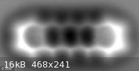
Single molecule, one million times smaller than a grain of sand, pictured for first time
Pretty cool.
http://tinyurl.com/knhsmt
"....
Scientists from IBM used an atomic force microscope (AFM) to reveal the chemical bonds within a molecule.
'This is the first time that all the atoms in a molecule have been imaged,' lead researcher Leo Gross said....."
Now if they only add color and better focus - I'd buy two 

pentacene
[Edited on 30-8-2009 by argyrium]
|
|
|
hissingnoise
International Hazard
    
Posts: 3940
Registered: 26-12-2002
Member Is Offline
Mood: Pulverulescent!
|
|
I think M Rothko beat them to it?
But seriously, it is mindblowing to know you're actually seeing the fused rings, soft-focus though they are. . .
[Edited on 30-8-2009 by hissingnoise]
|
|
|
Klute
International Hazard
    
Posts: 1378
Registered: 18-10-2006
Location: France
Member Is Offline
Mood: No Mood
|
|
Incredible.
I wonder how they fixed that single molecule of carbon monoxide at the exact point of a metallic pyramide.. How do you construct such nanomaterial at
one molecule close?
\"You can battle with a demon, you can embrace a demon; what the hell can you do with a fucking spiritual computer?\"
-Alice Parr
|
|
|
chemoleo
Biochemicus Energeticus
    
Posts: 3005
Registered: 23-7-2003
Location: England Germany
Member Is Offline
Mood: crystalline
|
|
I wonder though how this could be used for anything more advanced- for structures of chemical compounds - very difficult as they may not be planar,
and as soon as they stick off the plane, multiple rotational 'snapshots' need to be taken to get an idea of the overall structure. Also no atom
information is given with this method. With complex structures, such as large biological macromolecules (proteins, lipids, carbohydrates and their
infinite combinations), all you'll ever get is the surface, and focus will be an issue with showing a 3D molecule (unlike the 2D pentacene molecular
structure). And for getting the surface texture of complex molecules (albeit at a lower res) there are plenty of existing methods.
Not only that, for any more complex molecules such as proteins (where this would be of most profound use), there's still the issue of having a large
molecule sitting on a sticky plane (with no water, salt, buffer etc keeping them happy, stable, folded and active)- and almost certainly it will not
look like the molecule in solution. There'll be artefacts much much worse than with crystals of proteins (also worse than with electron microscopy)
Nonetheless, it is pretty cool!
Didn't they do this with gold atoms already btw?
[Edited on 1-9-2009 by chemoleo]
Never Stop to Begin, and Never Begin to Stop...
Tolerance is good. But not with the intolerant! (Wilhelm Busch)
|
|
|
psychokinetic
National Hazard
   
Posts: 558
Registered: 30-8-2009
Location: Nouveau Sheepelande.
Member Is Offline
Mood: Constantly missing equilibrium
|
|
Can't Berkley already see individual atoms?
“If Edison had a needle to find in a haystack, he would proceed at once with the diligence of the bee to examine straw after straw until he found
the object of his search.
I was a sorry witness of such doings, knowing that a little theory and calculation would have saved him ninety per cent of his labor.”
-Tesla
|
|
|
JohnWW
International Hazard
    
Posts: 2849
Registered: 27-7-2004
Location: New Zealand
Member Is Offline
Mood: No Mood
|
|
Yes, with an electron microscope, but only of conducting
materials like metals.
|
|
|
Ozone
International Hazard
    
Posts: 1269
Registered: 28-7-2005
Location: Good Olde USA
Member Is Offline
Mood: Integrated
|
|
IIRC they spelled out "IBM" with xenon (which are very large, for atoms) atoms :
http://www.fourmilab.ch/autofile/www/section2_84_14.html
http://www.nanooze.org/english/interviews/doneigler.html
Wish I could find my copy of Time Science:Matter, which has a nicer B/W of the image than I could find on the web. It suddenly occurs to me that media
format is part of the problem with relaying science to the masses. They go into how wonderful and mischeviously complex "extreme scientificky X thing"
is, but forget? to put the cool image in the article.
Result: very few care enough to read the article.
The picture says it all--put it to press (regardless of the extra cost), damnit.
[edit] Oh yes (re. pentacene), remember that atoms are not perfectly hard spheres. They are tiny nuclei surrounded at a (relatively) great (read
immense) distance by an electron cloud. The fact that it is not a peanut-shaped blob is pretty impressive to me (particularly considering the electron
delocalization in this system). I think that the picture was quite crisp. Quite cool.
Cheers,
O3
[Edited on 1-9-2009 by Ozone]
-Anyone who never made a mistake never tried anything new.
--Albert Einstein
|
|
|
Magpie
lab constructor
    
Posts: 5939
Registered: 1-11-2003
Location: USA
Member Is Offline
Mood: Chemistry: the subtle science.
|
|
I was awed by this picture. It is just fantastic to be able to see something that small. Thanks for posting this, argyrium.
And what a wonderful confirmation of the atomic theory and molecular bonding. Ever since the Greeks came up with the concept of atoms we have only
been able to gather inferential evidence. Now we can see them!
The single most important condition for a successful synthesis is good mixing - Nicodem
|
|
|
Sedit
International Hazard
    
Posts: 1939
Registered: 23-11-2008
Member Is Offline
Mood: Manic Expressive
|
|
I must ask the noob question of whats the difference between this picture and those imaged thru the use of a SEM? I admit I have not read the link yet
so bear with me.
Knowledge is useless to useless people...
"I see a lot of patterns in our behavior as a nation that parallel a lot of other historical processes. The fall of Rome, the fall of Germany — the
fall of the ruling country, the people who think they can do whatever they want without anybody else's consent. I've seen this story
before."~Maynard James Keenan
|
|
|
Ozone
International Hazard
    
Posts: 1269
Registered: 28-7-2005
Location: Good Olde USA
Member Is Offline
Mood: Integrated
|
|
Looks like this to me:
0. AFM with a single molecule (CO) probe.
1. Carbon is much smaller than the Xe or metal atoms that they have been pushing around.
2. The connectivity (and planar nature of this particular system) of the atoms, and, it looks like, the pi e- delocalization in an aromatic system
were unambiguously imaged (Interesting how the signals are so much greater on the "ends"; I wonder if that is real or if it is an artifact).
I need to get the paper (the blurb is in C&EN, this week).
Heady stuff,
O3
-Anyone who never made a mistake never tried anything new.
--Albert Einstein
|
|
|
watson.fawkes
International Hazard
    
Posts: 2793
Registered: 16-8-2008
Member Is Offline
Mood: No Mood
|
|
Quote: Originally posted by Ozone  | | the pi e- delocalization in an aromatic system were unambiguously imaged (Interesting how the signals are so much greater on the "ends"; I wonder if
that is real or if it is an artifact). |
Sure looks real to me. Think of Gauss's Law applied at the
microscale. Excess charge has migrated to the surface of a conductor. In this case the surface has two ends. I even suspect the difference in
intensity between the two ends is real, given that the charge distribution is almost-certainly quantized, yielding local minima and metastable states.
|
|
|
psychokinetic
National Hazard
   
Posts: 558
Registered: 30-8-2009
Location: Nouveau Sheepelande.
Member Is Offline
Mood: Constantly missing equilibrium
|
|
Here we go: Link to video of the microscope and its use
“If Edison had a needle to find in a haystack, he would proceed at once with the diligence of the bee to examine straw after straw until he found
the object of his search.
I was a sorry witness of such doings, knowing that a little theory and calculation would have saved him ninety per cent of his labor.”
-Tesla
|
|
|
turd
National Hazard
   
Posts: 800
Registered: 5-3-2006
Member Is Offline
Mood: No Mood
|
|
Um, no. You're probably thinking about a TEM or STEM microscope and these work with insulators as well. Also, while the lateral resolution is smaller
than an atom, technically what you're seeing is a column of atoms. You're not going to mount a single atom layer into your sample holder. And, while
the images look very neat, interpretation is not as easy as you might believe at first. That's why the HRTEM guys do lots of simulation.
OTOH, single atoms of solid state compounds have been pictured with AFM in the 1980s, but that's much easier than single molecules since, with a
typical AFM, you would just move those around or destroy them. While the pictures produced with AFM look great, its actually a quite annoying method.
The tip quickly gets dirty and makes the image quality decrease. Lots of work until you get the one picture you want!
PS: If I was rich, a 200 kV TECNAI TEM would be one of the first things I'd buy. This is just too cool of an instrument.
|
|
|
psychokinetic
National Hazard
   
Posts: 558
Registered: 30-8-2009
Location: Nouveau Sheepelande.
Member Is Offline
Mood: Constantly missing equilibrium
|
|
Quote: Originally posted by turd  |
PS: If I was rich, a 200 kV TECNAI TEM would be one of the first things I'd buy. This is just too cool of an instrument. |
*drools*
“If Edison had a needle to find in a haystack, he would proceed at once with the diligence of the bee to examine straw after straw until he found
the object of his search.
I was a sorry witness of such doings, knowing that a little theory and calculation would have saved him ninety per cent of his labor.”
-Tesla
|
|
|
hinz
Hazard to Others
  
Posts: 200
Registered: 29-10-2004
Member Is Offline
Mood: No Mood
|
|
Quote: Originally posted by turd  |
PS: If I was rich, a 200 kV TECNAI TEM would be one of the first things I'd buy. This is just too cool of an instrument. |
Bah, TEM sample preparation is quite complex, at least if you want to see more advanced things than metal structures.
The grinding of the sample is quite time consuming, you can easily spend a half a day grinding your sample to a thickness of about 100nm (to get it
electron transparent) by using finer and finer grinding paper. And if you don´t pay attention, your sample gets flushed down the drain if the glue
mounting to the tripod fails. Or you grind to much and there´s no more sample
If your sample is brittle, like semiconductors, glass or ceramics, the sample also likes to break.
So if you need to prepare samples from these materials you´ll need a FIB (Focussed Ion Beam), which is also real expensive.
For SEM you´ll only need a sputtering/carbon coating machine
So if there is the alternative between SEM and TEM, I would always prefere the SEM and only use the TEM if the magnification is very important. IMHO a
SEM is much more universal and cheaper than a TEM.
[Edited on 1-9-2009 by hinz]
|
|
|
sparkgap
International Hazard
    
Posts: 1234
Registered: 16-1-2005
Location: not where you think
Member Is Offline
Mood: chaotropic
|
|
| Quote: | The Chemical Structure of a Molecule Resolved by Atomic Force Microscopy
Leo Gross, Fabian Mohn, Nikolaj Moll, Peter Liljeroth, Gerhard Meyer
Science,2009, 325 (5944), pp 1110–1114/i]
Abstract
Resolving individual atoms has always been the ultimate goal of surface microscopy. The scanning tunneling microscope images atomic-scale features on
surfaces, but resolving single atoms within an adsorbed molecule remains a great challenge because the tunneling current is primarily sensitive to the
local electron density of states close to the Fermi level. We demonstrate imaging of molecules with unprecedented atomic resolution by probing the
short-range chemical forces with use of noncontact atomic force microscopy. The key step is functionalizing the microscope’s tip apex with suitable,
atomically well-defined terminations, such as CO molecules. Our experimental findings are corroborated by ab initio density functional theory
calculations. Comparison with theory shows that Pauli repulsion is the source of the atomic resolution, whereas van der Waals and electrostatic forces
only add a diffuse attractive background. |
They have been playing with pentacene for a long time, after all...
sparky (~_~)
Attachment: pentapic.pdf (973kB)
This file has been downloaded 853 times
"What's UTFSE? I keep hearing about it, but I can't be arsed to search for the answer..."
|
|
|
turd
National Hazard
   
Posts: 800
Registered: 5-3-2006
Member Is Offline
Mood: No Mood
|
|
Quote: Originally posted by hinz  | | Bah, TEM sample preparation is quite complex, at least if you want to see more advanced things than metal structures. |
What could be easier than putting a drop of an emulsion on a carbon grid and dry it?
Of course you are right about the tediousness of preparing solid samples.
Quote: Originally posted by hinz  | If your sample is brittle, like semiconductors, glass or ceramics, the sample also likes to break.
So if you need to prepare samples from these materials you´ll need a FIB (Focussed Ion Beam), which is also real expensive.
|
Indeed, that's why next to most TEMs you'll have a SEM/FIB machine. To prepare samples. 
Quote: Originally posted by hinz  | | So if there is the alternative between SEM and TEM, I would always prefere the SEM and only use the TEM if the magnification is very important. IMHO a
SEM is much more universal and cheaper than a TEM. |
I think you completely miss the point of TEM: simultaneous imaging and diffraction.
|
|
|
Formatik
National Hazard
   
Posts: 927
Registered: 25-3-2008
Member Is Offline
Mood: equilibrium
|
|
Pretty amazing to see a molecule like this, and good to see some theory further verified.
|
|
|
franklyn
International Hazard
    
Posts: 3026
Registered: 30-5-2006
Location: Da Big Apple
Member Is Offline
Mood: No Mood
|
|
Honestly you're all going off the deep end here.
Nothing is being " seen ". Feeling around in the
dark is a better description. This is perhaps a
bit better technique than crystallography which
actually " sees " the shadow cast by molecules.
What is interesting is not the experimental
affirmation of postulated structure but what
can be learned at a size where quantum effects
become pronounced.
Because they are set unusually far apart , atoms
of uranium in its oxide have been imaged by
electron microscopy many years ago.
.
|
|
|
turd
National Hazard
   
Posts: 800
Registered: 5-3-2006
Member Is Offline
Mood: No Mood
|
|
Quote: Originally posted by franklyn  | | Honestly you're all going off the deep end here. Nothing is being " seen ". Feeling around in the dark is a better description. This is perhaps a bit
better technique than crystallography which actually " sees " the shadow cast by molecules. |
These two methods are so different in their application and their results that saying one or the other is better makes no sense at all.
Crystallography works on three dimensional crystalline (more or less) compounds and has an insanely high accuracy, whereas AFM is purely a surface
method. Try solving the structure of a protein, a metal-organic compound or even an inorganic solid-state compound with AFM or "picturing" the
structure of a single molecule or mono-layer with XRD. Good luck.
Also I highly object the use of the word "shadow" to describe diffraction. There's a point when simplification for the masses becomes
oversimplification and this is way past it. A shadow cast by the sun is a projection of 3-dimensional space on 2-dimensional space, therefore throwing
away 3d information. What you measure in crystallography is the Fourier transform of the electron density, but only intensity without the phase data.
So in a way, if you wish to call it so, the "shadow" of the Fourier transform. But, in practically all cases, the missing phase data can be recovered
by the fact that the crystal structure must be physically sound, i.e. no negative electron density and sharp peaks which are not too close. (There
are some freak structures where the diffraction image is not unambiguous, a fact which is known since the dawn of X-ray diffraction. But this is not
your common boring organic molecule.)
Quote: Originally posted by franklyn  | | Because they are set unusually far apart , atoms of uranium in its oxide have been imaged by electron microscopy many years ago.
|
The difference between TEM/SEM/AFM of bulk and single molecules has already been discussed above. All these methods measure different things. Read the
thread.
|
|
|
psychokinetic
National Hazard
   
Posts: 558
Registered: 30-8-2009
Location: Nouveau Sheepelande.
Member Is Offline
Mood: Constantly missing equilibrium
|
|
Feeling around in the dark makes sense, but then the same could be said for many forms of imaging...not only in chemistry I suppose.
“If Edison had a needle to find in a haystack, he would proceed at once with the diligence of the bee to examine straw after straw until he found
the object of his search.
I was a sorry witness of such doings, knowing that a little theory and calculation would have saved him ninety per cent of his labor.”
-Tesla
|
|
|
franklyn
International Hazard
    
Posts: 3026
Registered: 30-5-2006
Location: Da Big Apple
Member Is Offline
Mood: No Mood
|
|
@ turd
My "shadow" metaphor if taken literally is not descriptive of the methodology as
you have detailed somewhat. You are welcome to provide a more accurate descriptor.
At large scale , computer axial tomography ( CAT ) scanning unlike optical polaroids
similarly displays images produced as interpretations from the gathered data.
" Seeing " unless it is itself just an abstract reference such as "viewing " " picturing "
" imaging " or " resolving " is just as misrepresentive of what is in essence remote sensing
of nano scale objects and their depiction as a graph - what prompted my comment.
" All these methods measure different things " ,
- gee I hope not , Robin Williams said it best " reality , what a concept "
and Lilly Tomlin " what's reality anyway , nothing but a collective hunch "
.
|
|
|
thaflyemcee
Harmless

Posts: 12
Registered: 14-8-2009
Member Is Offline
Mood: No Mood
|
|
There are images of orbitals in W and Si observed by AFM a few years back. It relies on some neat tricks, like getting the sample to image the tip,
but it's totally valid. I'll post the paper once I find it again.
Before anyone jumps on me for "orbitals are mathematical constructs", let me remind you that on the timescale on which AFM image is acquired, we are
ultimately looking at a map of where the tip was repelled by the surface on average, which allows the geometry of allowed electron
positions (i.e., an orbital) to be imaged.
|
|
|
Ozone
International Hazard
    
Posts: 1269
Registered: 28-7-2005
Location: Good Olde USA
Member Is Offline
Mood: Integrated
|
|
"Fractal molecule bacteria!"
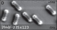
I have attached a shot of the other picture (on ~60A scale) which shows many pentacene molecules. Note that they all exhibit greater electron density
at the ends.
It verifies theoretical predictions and it does so with very nice "science fair appeal".
Now, I want to see anti-aromaticity . .
Cheers,
O3

-Anyone who never made a mistake never tried anything new.
--Albert Einstein
|
|
|
JohnWW
International Hazard
    
Posts: 2849
Registered: 27-7-2004
Location: New Zealand
Member Is Offline
Mood: No Mood
|
|
Quote: Originally posted by Ozone  | I have attached a shot of the other picture (on ~60A scale) which shows many pentacene molecules. Note that they all exhibit greater electron density
at the ends. It verifies theoretical predictions and it does so with very nice "science fair appeal".
Now, I want to see anti-aromaticity. |
The greater electron density observed in the pi orbitals at the ends of pentacene molecules would be explainable by their mutual electrostatic
repulsion.
"Anti-aromaticity"? Perhaps someone could write to that AFM micrographer, to suggest that he tries the same technique on molecules of
cyclooctatetraene, and on suitably stable derivatives of cyclobutadiene.
|
|
|
| Pages:
1
2 |